Standard seborrheic keratosis removal options include cryotherapy, curettage with electrosurgery, shave excision, Er:YAG and CO2 lasers, and topical 40% hydrogen peroxide (HP40) or nitric–zinc solutions, sometimes in combination. The most important rule: have a professional assess the lesion first, with dermoscopy or biopsy when it is changing, inflamed or suspicious. Size, thickness, location and skin tone then guide the method. We offer in-clinic cryotherapy and lesion removal.
TL;DR:
- Changing, inflamed, bleeding, or suspicious lesions need professional assessment, often with dermoscopy or biopsy, before removal so tissue remains available for diagnosis.
- HP40 left 65% of facial lesions clear or nearly clear, compared with 46% on the trunk and 38% on the extremities.
- Cryotherapy suits modest body lesions, but thick growths may need another session, and freezing can leave lighter patches, especially on darker skin.
- Shave excision preserves tissue for laboratory review when a lesion changes, bleeds, or looks atypical; curettage with electrosurgery is often used for thicker raised growths.
- Treating one lesion first lets you assess healing, scarring, and pigment changes before deciding whether to remove additional growths using the same method.
Table of Contents
- Which seborrheic keratosis removal options exist, and what is each best for?
- How does cryotherapy work, and when should you choose it?
- What happens with curettage and electrosurgery?
- When is shave excision the right choice?
- How do Er:YAG and CO2 lasers compare for removal?
- What do trials say about topical HP40 and nitric–zinc?
- Why do clinicians combine methods, and what is emerging?
- How do you choose the right method for your lesion?
- What should you expect before, during and after a removal visit?
- What are the risks, and what cosmetic results are realistic?
- Our perspective on assessment and safe removal
- Ready for a professional assessment and removal?
- FAQ
- Sources
Which seborrheic keratosis removal options exist, and what is each best for?
Think of this as a map. Each method gets its own section below, but this shortlist helps you see where you probably fit.
- Cryotherapy: liquid nitrogen freezes the lesion, which blisters, crusts and falls off over a couple of weeks. It suits many trunk and limb lesions and quick, in-office visits.
- Curettage with electrosurgery: the lesion is scraped off under local anesthetic and the base is cauterized. It works well for thicker lesions and heals into a flat, shallow wound.
- Shave excision: a blade removes the lesion at skin level, and the tissue can go to a lab. It is the go-to when a lesion looks unusual or bleeds.
- Er:YAG and CO2 lasers: light energy vaporizes the lesion layer by layer. They fit single lesions where cosmetic appearance is the top priority, and they depend heavily on operator skill.
- Topical HP40 and nitric–zinc: applied in repeated sessions with no cutting. They suit thinner lesions, especially on the face, for people who prefer a non-surgical route.
- Combination approaches: debulk first, then treat the base. These are used when one method alone is unlikely to clear a stubborn lesion.
In general, thin facial lesions lean toward gentler options such as topical therapy or a carefully chosen laser, while thicker trunk lesions often go to cryotherapy or scrape-and-cauterize methods. One reminder before anything else: a clinical review of treatment options describes these growths as benign, but they can imitate other skin conditions, so a professional should look first.
How does cryotherapy work, and when should you choose it?
Cryotherapy is the workhorse of seborrheic keratosis removal. A clinician applies liquid nitrogen with a spray device or a cotton-tipped applicator, freezing the lesion and a thin rim of surrounding skin. You feel a sharp cold and a brief sting or burning sensation that soon passes. Larger or thicker lesions may need a longer freeze or a second pass.
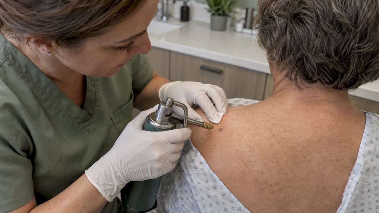
Dermatology references describe freezing as one of the classical in-office approaches, alongside curettage, as in this clinical overview from a university dermatology department. The same department notes that some methods require more than one treatment. A review of treatment options likewise lists cryotherapy among the classical approaches, and it is commonly used for lesions on the trunk and limbs.
What healing looks like:
- Early on: the area turns red and may swell slightly.
- During healing: a blister can form, sometimes a blood-tinged one.
- As it heals: the blister dries into a crust that falls off on its own.
- After the crust drops: the new skin underneath is pink and gradually fades.
Wound care is simple. Keep the area clean, apply a thin layer of petroleum jelly if your clinician suggests it, and never pick the crust. Sun protection matters afterward because fresh skin pigments easily.
The main trade-offs:
- Pigment loss: freezing can damage pigment cells, so lighter patches may appear, and this is a bigger concern on darker skin tones.
- Incomplete removal: thick lesions can be too deep for a single freeze, and a lesion may need a repeat session.
- Less suited to the delicate face: a gentler method may be preferable near the eyes or on thin facial skin.
Pro Tip: If you have darker skin, ask your clinician how long they plan to freeze and whether a test lesion in a less visible area makes sense before treating anything on your face.
Choose cryotherapy when you have a few modest lesions on the body, want a fast visit, and are comfortable with a blister and crust phase. Pick an alternative when the lesion is thick, in a high-visibility spot, or looks suspicious and needs tissue examined. If you are considering this route, our skin lesion removal page explains how we handle in-clinic cryotherapy for benign lesions.
What happens with curettage and electrosurgery?
Curettage with electrosurgery is the scrape-and-cauterize approach. Because seborrheic keratoses sit on top of the skin like a stuck-on wax drop, a sharp spoon-shaped instrument called a curette can lift them away cleanly, which is why this method is a classic choice. A review of current options lists curettage and electrodesiccation among the available treatments.
The steps look like this:
- Numbing: the clinician injects a small amount of local anesthetic, which stings briefly and then the area goes numb.
- Scraping: the curette removes the lesion.
- Electrosurgery: a fine electrical tip seals small blood vessels and treats the base so bleeding stops and residual cells are destroyed.
- Dressing: a small bandage or ointment goes on, and you leave with written aftercare steps.
Compared with cryotherapy, the experience trades one set of inconveniences for another. The anesthetic injection is the uncomfortable part, but the procedure itself is painless and there is usually no big blister phase. Many patients find the aftercare simpler, since the area forms a thin scab instead of a blister and crust. Others prefer freezing because it avoids needles. Neither is universally better, so personal preference matters.
Clinicians favor this method for thicker or raised lesions, for lesions that cryotherapy might not fully clear, and when they want a specimen. A curetted lesion can be sent for examination if anything looks atypical, so a biopsy is simple to arrange at the same visit.
Cosmetic considerations are worth knowing:
- Scarring: removal goes below the surface, so a flat, slightly pale or pink mark can remain, particularly if the area is picked or sun-exposed during healing.
- Pigment changes: lighter or darker patches are possible, as with most destructive methods.
- Healing time: expect a scab during healing, and the pink area can take longer to blend in.
If your lesion is large or has been irritated, a clinician may suggest this method precisely because it allows more control over depth.
When is shave excision the right choice?
Shave excision is as much about diagnosis as removal. Using a small blade, the clinician slices the lesion off at skin level, usually after numbing the area. The sample can then go to a pathology lab, so you get an answer about what the lesion actually was along with the removal.
This matters because seborrheic keratoses can look like other growths. The Canadian public-health guidance on skin cancer stresses having changing or suspicious lesions evaluated and warns that removing a lesion yourself can destroy the tissue a pathologist needs for an accurate diagnosis.
Shave excision tends to be chosen when:
- The lesion looks atypical: uneven color, irregular borders, or a texture that does not fit the classic stuck-on appearance.
- It is inflamed or bleeding: irritated lesions are harder to judge visually and may need tissue confirmation.
- It is changing: growth, darkening or new symptoms deserve laboratory review.
- You want a single-visit solution: the lesion is removed and tissue is preserved in one step.
Recovery looks a lot like curettage. A small shallow wound forms, is covered, and scabs over within days. Healing takes time, with the pink area fading gradually. Scarring risk is low for superficial shaves but rises if the blade goes deeper, and light or dark pigment changes can still occur.
If a pathology report comes back, ask for a follow-up conversation rather than just a notification. A benign result is reassuring, and an unexpected one means you have caught something early.
How do Er:YAG and CO2 lasers compare for removal?
Lasers remove seborrheic keratoses by vaporizing thin layers of tissue with focused light. Two types come up most: the erbium-doped YAG (Er:YAG) laser and the carbon dioxide (CO2) laser.
Er:YAG is gentler, with shallow penetration and less heat spreading into nearby skin. A clinical review of seborrheic keratosis reports that in a small comparative trial Er:YAG laser achieved higher clearance than cryotherapy, and notes that cryotherapy was associated with more hyperpigmentation in those cited studies. CO2 lasers are also effective but deliver more heat, which raises the chance of scarring and pigment change. The 2022 update on treatment options highlights pigmentary change and scarring as risks with some of these techniques.
Keep the evidence in perspective. The comparative trials are small, so treat them as encouraging rather than conclusive. Results also depend heavily on the person holding the laser, so experience with seborrheic keratosis and with your skin tone matters.
Lasers are practical when:
- Appearance is the top priority: a single visible lesion on the face or neck is a good candidate.
- You want precise depth control: the clinician can remove the lesion layer by layer.
- Other methods are poor fits: for example, when freezing risks pigment loss in your skin type.
The limitations are real:
- Cost: laser sessions typically cost more than cryotherapy.
- Limited large trials: the evidence base is thin compared with older methods.
- Availability: not every clinic offers both laser types.
- Operator dependence: technique strongly influences outcomes.
If you are weighing lasers, ask how many seborrheic keratoses the provider treats with the device, what settings they use for your skin tone, and what the plan is if the lesion does not fully clear.
What do trials say about topical HP40 and nitric–zinc?
Topical therapy is the non-surgical option, and the evidence has grown in recent years. The two main candidates are a 40% hydrogen peroxide solution (HP40) and a nitric–zinc complex.
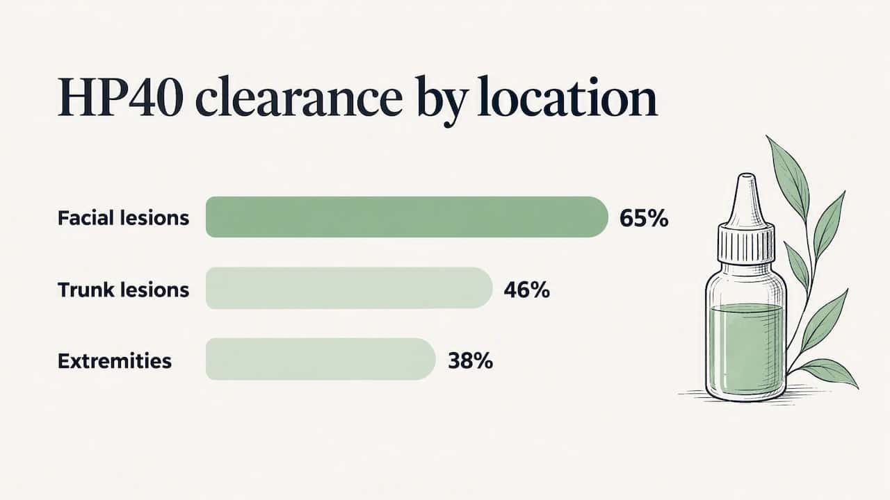
For HP40, a Cleveland Clinic overview describes a pooled analysis of 937 patients in which facial lesions did notably better than lesions elsewhere on the body. Typical reactions were mild redness, stinging and swelling that resolved within about a week.
A pooled analysis of 937 patients found 65% of facial seborrheic keratoses clear or near-clear with HP40, versus 46% on the trunk and 38% on the extremities (Cleveland Clinic). Facial skin is thinner, which appears to help the solution do its work.
A review of recent advances also describes HP40 as a promising alternative, particularly for facial lesions. For nitric–zinc complex, the clinical review of treatment options notes it has been investigated recently, and cited series reported clearance in many treated lesions after a few applications with minimal discomfort. As with lasers, these reports are small, so they are encouraging but not definitive.
Who tends to be a good candidate for topical therapy:
- Thin facial lesions: where HP40 performs best.
- People wary of cutting or freezing: topical care involves no blade or anesthetic.
- Patients who accept multiple sessions: repeat applications are usually needed.
- Those with lesions near sensitive areas: where destructive methods feel riskier.
The trade-off is time and thoroughness. Thicker trunk lesions respond less often, and you may need several visits. Topical treatment is applied in a clinical setting rather than at home, so ask your provider who administers it and how many sessions to expect.
Why do clinicians combine methods, and what is emerging?
Sometimes one method is not enough. A thick, stubborn lesion can resist a single freeze, and a lesion that is too raised for a laser can be debulked first. The most common combination is shaving or curetting the bulk of the lesion, then using electrosurgery to coagulate the base and stop bleeding. This pairs the speed of mechanical removal with the control of cautery.
Other combinations mix a mechanical step with cryotherapy for the base, or follow a first treatment with a topical session if a thin residual patch remains. The point is to match the tool to each layer of the lesion instead of forcing one method to do everything.
Reviews also mention emerging techniques, including nanosecond-pulsed electric field treatment and a range of laser types. The 2022 update on pathophysiology and treatment covers the wider landscape of options. The caveat is that large head-to-head trials are lacking, so no newer method has displaced the standard ones.
When single methods are unlikely to clear a lesion, bring it up directly. Ask whether a staged or combined plan makes sense, how many visits it involves, and what the expected cosmetic result is. A thoughtful provider will explain why they chose each step.
How do you choose the right method for your lesion?
Start with the features that drive the decision:
- Size and thickness: thin, flat lesions suit topical therapy or light freezing; thick, raised ones lean toward curettage or combinations.
- Location: the face favors gentler options; the trunk tolerates freezing and scraping more readily.
- Skin tone: darker skin has a higher risk of pigment change with freezing and some lasers.
- Number of lesions: many lesions may call for a staged plan.
- Cosmetic priority: visible spots may justify a more precise, costlier option.
- Healing preference: blister and crust, or a small scabbed wound, or no wound at all.
At your consultation, work through these questions:
- Was dermoscopy used? A close look with a dermatoscope helps tell seborrheic keratosis from lookalikes, as the review of recent advances notes for pigmented lesions.
- Is a biopsy recommended? Ask why or why not, especially if the lesion has changed.
- What downtime should I expect? Get a realistic healing timeline.
- What is the scarring and pigment-change risk for my skin tone?
- How many sessions will I need? Some methods require repeat visits.
- What will it cost, and is it per lesion or per session?
Pro Tip: If you have several lesions, treat one first. It shows you how your skin heals and lets you judge the cosmetic result before committing to the rest.
This one-lesion-first approach also gives you a chance to reconsider. If the healing is slower or the pigment shift is more noticeable than you hoped, you can switch methods for the next spot.
What should you expect before, during and after a removal visit?
Knowing the flow takes much of the anxiety out of the appointment.
Before:
- Assessment: the clinician examines the lesion, usually with dermoscopy, and may photograph it for your record.
- Discussion: you talk through options, risks and expected healing.
- Consent: you agree to a specific method, and a biopsy is arranged if indicated.
During:
- Numbing if needed: a local anesthetic for curettage or shave excision; none for most freezing.
- The procedure: the time depends on the method and the lesion.
- Specimen handling: if tissue is going to a lab, it is labeled and sent, and you are told when to expect results.
After:
- Cleaning and dressing: wash gently and keep the area covered or moisturized as advised.
- Sun protection: shield the area from sun while it heals to reduce pigment changes.
- Hands off: do not pick scabs or crusts.
- Expected timeline: blistering and crusting as the area heals after freezing, or a thin scab as it heals after scraping or shaving.
- Signs of infection: spreading redness, warmth, pus, increasing pain or fever warrant a call to your clinic.
Plan a follow-up if the lesion does not clear, grows back, or looks different. Canadian clinical nursing guidance on skin conditions, for example, notes that scarring and secondary infection are recognized complications, which is why aftercare and follow-up matter.
What are the risks, and what cosmetic results are realistic?
Most seborrheic keratosis removal methods are safe in trained hands, but none are risk-free. Mild redness, swelling and tenderness are common and temporary. The bigger questions are lasting marks.
- Pigment changes: lighter or darker patches can follow freezing, electrosurgery and lasers, and are more noticeable on darker skin. The 2022 treatment update flags pigmentary change as a risk with some techniques.
- Scarring: deeper or heat-based methods carry a higher scar risk, with CO2 laser and aggressive curettage at the upper end.
- Facial lesions: in trial data, facial lesions responded well to topical HP40, with milder reactions than destructive options, which makes it attractive where appearance matters most.
- Recurrence: the removed lesion usually does not return, but new seborrheic keratoses can appear nearby, and an incompletely cleared lesion may regrow.
If something returns or looks different, tell your clinician. They may retreat with the same method or suggest a combined approach, and any change in a lesion deserves a fresh look.
Our perspective on assessment and safe removal
We specialize in non-invasive skincare, and we offer cryotherapy and benign lesion removal in a private, personalized setting. Our workflow starts with a careful look at the lesion, including dermoscopy, followed by a conversation about options and informed consent before anything is done. We then recommend a method that fits your lesion and skin, give clear aftercare instructions, and plan a follow-up so healing does not go unchecked. Safety comes first: if a lesion is changing, bleeding or simply looks wrong to you, we urge a professional assessment before any removal.
— Andy
Ready for a professional assessment and removal?
If you have a lesion you want looked at or removed, we can help you take it step by step. Our cryotherapy for benign skin lesions is priced at 25-40 CAD per lesion, with an appointment fee of 75 CAD.
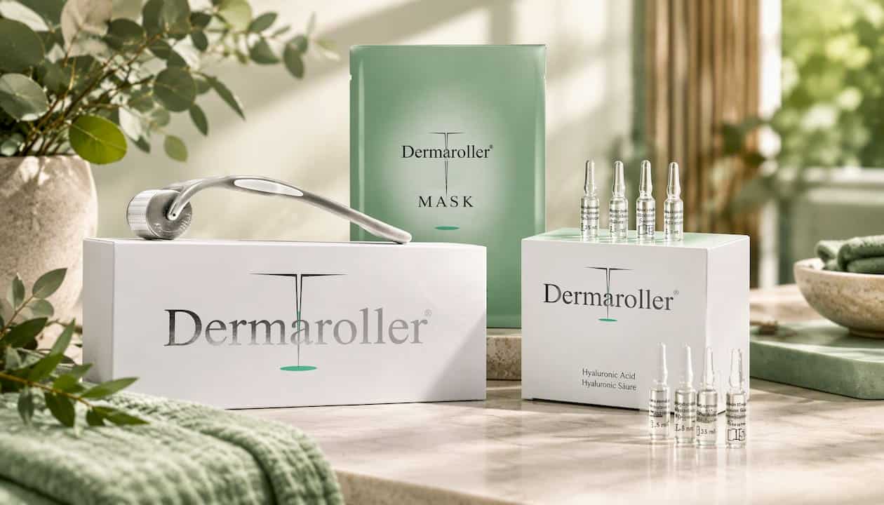
After treatment, supporting your skin can help it look its best. These resources may be useful:
- Collagen Induction Therapy / Micro-Needling: a professional treatment at 350 CAD to support skin texture once lesions have healed.
- Ultimate Dermaroller® Home Care Set: a home-care option for ongoing skin support.
- Dermaroller® Home Care and Dermaroller® Roller: companion products for your routine.
Your next steps are simple: book an appointment, get your lesion assessed, choose a method with your clinician, and plan your follow-up. You can reach us through our website.
This article is general information, not a substitute for advice from a qualified doctor. Consult a qualified healthcare professional about your own circumstances before acting on anything here.
FAQ
How can seborrheic keratosis be removed?
Seborrheic keratosis can be removed in a clinic by freezing with liquid nitrogen, scraping with a curette plus electrosurgery, shaving, or laser vaporization. Topical options such as HP40 are also used, especially for facial lesions. A clinician chooses based on the lesion’s size, thickness and location, and your skin tone.
What is seborrheic keratosis around the bra line?
Seborrheic keratoses often appear where skin rubs against clothing, including the bra line, and the friction can make them irritated or inflamed. They remain benign, but irritation is a good reason to have a clinician look and, if needed, remove them. Because irritated lesions can look unusual, professional assessment is wise.
Can kids have seborrheic keratosis?
Seborrheic keratoses are far more common in adults, and incidence rises with age and sun exposure, according to a review of recent advances. A dark or changing spot on a child should be seen by a clinician rather than assumed to be one.
Can I use a wart remover to remove seborrheic keratosis?
Over-the-counter wart removers are not designed for seborrheic keratosis and can irritate or scar the skin. Removing a lesion yourself can also destroy tissue needed for diagnosis, which Canadian public-health guidance cautions against. Have a professional assess it first.
Sources
- Seborrheic Keratosis: What Is It, Causes, Risks & Treatment
- Seborrheic Keratoses – The Most Common Benign Skin Tumor of Humans. Clinical presentation and an update on pathogenesis and treatment options
- Canada